PROFESSIONAL EYE EXAMINATION
Historically, an eye examination was nothing more than flipping lenses before the eye by the optician and took no more than a few minutes to do the job. No information on the health condition of the eye, whether the eyes are working together or even if the prescription was right. There was no formal training for opticians and therefore absence of practice standards until some 20 years ago when the government started to regulate the profession in order to protect the public from low standards of optical service.
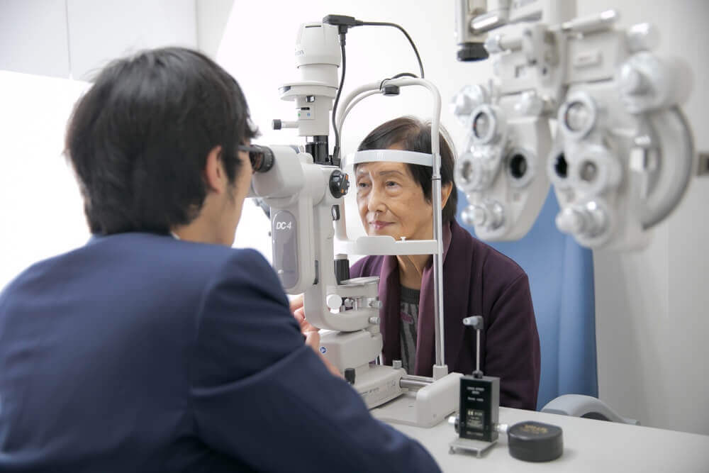
Today, Optometry is a professional study in the university and Optometrists are your primary eye care providers. Together with advanced equipment, optometrists are able to provide a comprehensive eye examination for everyone.
A routine eye exam includes, but not limited to, the following procedures:
- History taking and understanding of visual problems
- Assessment of visual functions (vision, visual acuity, visual field, colour vision, etc.)
- Refraction (finding your spectacle prescription)
- Binocular coordination (muscle balance, stereopsis, squint, lazy eyes, etc.)
- Ocular health assessment (glaucoma, cataract, retinal conditions, etc.), particularly when other findings are contradictory.
- Diagnosis and treatment of visual anomalies


OUR SERVICES
(1) Primary Eye Care Examination (Comprehensive Eye Examination)
A comprehensive eye examination comprises of 4 major test components:
1.Medical history taking and complaints
- To understand why a patient is seeking an eye examination and what are the major complaints. Individual and family medical and ocular histories are explored for any possible link to the complaints.
2.Visual functions tests
- Vision / visual acuity
- Contrast sensitivity
- Refraction (sight test) for short-sightedness, long-sightedness, astigmatism or presbyopia (aging sight)
- Binocular vision: muscle motility, focusing ability, squint, amblyopia (lazy eye) and stereopsis (depth perception)
- Pupil reflex
- Visual field
- Colour blindness
3.Ocular health tests
- Intraocular pressure
- Anterior and posterior segments: eyelid and annex, tears, conjunctiva, cornea, iris, crystalline lens, vitreous, retina and macula
- Dry eye
- Contact lens related complications
4.Case Analysis and management
- Analysis and explanation of clinical test findings
- Suggestion of appropriate actions or management plans
- Abnormal conditions, when suspected, will be referred to appropriate medical specialties
- Common referrals include:
- Glaucoma
- Cataract
- Floaters
- Macular degeneration
- Retinal holes, tears or detachments
- Diabetic retinopathy
- Retinal blood vessel occlusion
- Brain tumour
- Hypertensive retinopathy
Primary eye care examination not only ensures that you have clear and comfortable vision but also maintains that your visual system stays healthy and functional: a key to preventive eye care.

(2) Visual Functions Test
Visual functions are the ability of the various components of the eye to work together for us to perceive things around us. They comprehend clear single vision, colour, light contrast, movement, shape, depth, etc.
Visual functions tests include:
- Vision / visual acuity
- Contrast sensitivity
- Refraction (sight test) for short-sightedness, long-sightedness, astigmatism or presbyopia (aging sight)
- Binocular vision: muscle motility, focusing ability, squint, amblyopia (lazy eye) and stereopsis (depth perception)
- Pupil reflex
- Visual field
- Colour blindness

(3) Ocular Health Examination
Ocular Health Examination
Many sight threatening eye diseases may not have any symptoms in the early stages and our vision may still be fine. Ocular health examination is therefore vital in making sure that our visual system continues to function healthily.
Ocular health tests include the following:
- Intraocular pressure
- Anterior segment: eyelid and annex, tears, conjunctiva, cornea, iris, crystalline lens
- Posterior segments: vitreous, retina, macula
- Dry eye
- Contact lens related complications
Test results will be analyzed and explained to the patient and appropriate action or management plan will be suggested.
Abnormal conditions, when suspected, will be referred to appropriate medical disciplines。
Common referrals include:
- Glaucoma
- Cataract
- Floaters
- Macular degeneration
- Retinal holes, tears or detachments
- Retinal blood vessel occlusion
- Brain tumour
- Hypertensive retinopathy
- Diabetic retinopathy

(4) Dry Eye Assessment
Dry Eye Assessment
Dry Eye Disease is one the most common eye diseases worldwide.
Dry Eye Symptoms include:
- Dryness
- Grittiness
- Foreign body sensation
- Red eyes
- Sore eyes
- Photophobic
- Tearing
- Discharge
- Swelling and heavy eyelids
- Transient blurred vision
Dry Eye Examination includes assessments of:
- Ocular Surface Disease Index (OSDI) Questionnaire
- Tears meniscus height
- Tears volume
- Tears break up time
- Fluorescein / Lissamine Green staining of cornea and conjunctiva
- Meibomian gland function / dysfunction
- Ocular surface cell damage
- Tears lipid quality
- Tears pH-values


(5) Children’s Vision Examination
Children’s Vision Examination
Since birth, the eye starts to develop according to age, heredity, health conditions and environmental influences.
A comprehensive eye examination can start at the age of 3 and latest before the age of 6 as this is the critical period for vision development. Refractive errors, lazy eyes, abnormal eye conditions can be detected, corrected, managed and treated early enough to avoid permanent damage to vision. An eye examination as early as 6 months can also be conducted to rule out unusually high refractive errors, eye muscle incoordinations or congenital conditions such as cataract.
Common children’s vision problems include:
- Blurred vision: short-sightedness, long-sightedness, astigmatism
- Muscle imbalance between the two eyes: squint (cross-eye), poor depth perception.
- Weakness in accommodation: poor focus at near
- Learning difficulty
Lazy eyes or amblyopia, if diagnosed and treated before the age of 8, the prognosis can be very satisfactory. Beyond this critical period, it might be extremely difficult to restore good vision.
Children’s vision examination is no different from a primary eye care examination for adults except that a cycloplegic refraction test (using eyedrops) is necessary to confirm a true refractive error by eliminating a possible misdiagnosis of pseudo-myopia caused by overacting eye muscles.

(6) Myopia Management for Children
According to studies from the School of Optometry at the Hong Kong Polytechnic University, the prevalence of myopia (short-sightedness) for different age groups is shown below:
- Age 6 – 7 : 30%
- Age 10-11: 60%
- Age 16-17: 74%
- Age 19-39: 71%
It can easily be seen that the problem of myopia is very serious in Hong Kong.
According to other studies, currently one third of the world population is myopic and by Year 2050, half of the population will be myopic, equivalent to 5 billion people worldwide. Of these people, 1 billion of them will be of high myopia.
Myopia will become the most common cause of blindness.
More and more evidence has shown that myopia, high or low, will increase the risk of visual impairments and is higher for higher myopia. Myopia results in an elongation of the eye which causes the retinal and choroidal tissues to become thinner leading to a higher chance of retinal detachment and myopic maculopathy (disease of the macula). High myopia may also increase the incidence of cataract and glaucoma.
Researchers from a sample of 21,000 patients were able to demonstrate that for every increase of 1.00 dioptre in myopia, the prevalence of myopic maculopathy would increase by 67%.
Myopia management aims to control or slow down the progression of myopia by means of different methods or procedures in order to prevent future visual loss or blindness.
Therefore, myopia prevention and control are much more important than just correcting for it. In other words, when a child starts to develop and/or progress fast in myopia, myopia management strategy should be considered, in addition to a proper eye examination.
One or more of the following means can be considered for myopia control:
- D.I.M.S. optical defocus spectacle lenses (MyoSmart)
- Other optical defocus spectacle lenses (MyoVision)
- Progressive spectacle lenses (Myopilux)
- DISC-1 Day daily disposable soft contact lenses
- Other optical defocus daily disposable soft contact lenses (MiSight)
- Orthokeratology
- Atropine eye drops
(NB:This centre does not provide atropine eyedrops)

(7) Orthokeratology (Ortho-K)
Orthokeratology (Ortho-K)
Orthokeratology is the application of specially designed gas permeable contact lenses to gradually reshape the central and peripheral cornea for the correction of short-sightedness and control of myopia progression.
Orthokeratology lens is designed based on a combination of advanced ophthalmic instrumentation and precision computer technology together with the professional knowledge and experience of the Ortho-K practitioner.
According to studies from the Hong Kong Polytechnic University and others, Orthokeratology slows down myopia progression by about 50% on average.
Safe Ortho-K requires:
- A comprehensive eye exam with special attention to the health of the cornea, conjunctiva and tears.
- Strict compliance with hygiene and lens care regimen by parents and children
- A thorough understanding of the relationship between hygiene and contact lens complications.
- Regular follow up exams
In addition to regular contact lens instruments, special equipment such as a corneal topographer and an axial length scan are essential in monitoring changes and results.
Depending on the individual, Ortho-K may show its optical effect from a few hours to a few days or weeks and optimal reshaping effects can usually be achieved with one to three pairs of lenses. The wearer will then continue with the final retainer lenses during sleep for clear vision the following day.
Orthokeratology is suitable for people of all ages, especially young children during their growth stages, as long as they meet the refractive and health requirements. The safety of Ortho-K lenses is no different from any other contact lenses provided proper fitting, hygiene, lens care and follow up are closely observed.


(8) MIYOSMART Myopia Control Spectacle Lens
MiYOSMART is an innovative spectacle lens for myopia control developed by Hoya together with its partner, The Hong Kong Polytechnic University.Based on a two-year clinical trial results, MiYOSMART is proven to slow down myopia progression by up to 59% and halt myopia progression by 21.5% with its award-winning D.I.M.S. (Defocus Incorporated Multiple Segments) technology.
MiYOSMART with D.I.M.S. technology is comprised of a central optical zone for correcting refractive error and multiple defocus segments evenly surrounding the central zone (extending to the mid-periphery) of the lens to control myopia progression. This provides clear vision and myopic defocus simultaneously at all viewing distances.
MiYOSMART is made from Polycarbonate 1.59 material which provides good impact resistance and UV protection and is therefore safe to use for children.
In 2018, the MiYOSMART lens with D.I.M.S. technology was awarded Grand Prize, Grand Award and Special Gold Medal at the 46th International Exhibition of Inventions of Geneva, Switzerland.
(Source: HOYA)


(9) DISC-1 Day Myopia Control Soft Contact Lens
DISC-1 Day Myopia Control Soft Contact Lens
Myopia or shortsightedness occurs when the eyeball is too long relative to the focusing power of the eye. Light focuses in front of the retina rather than on it, making distant objects blurry.

The DISC lens, developed by the PolyU, is a multi-zone soft contact lens that provides clear vision and at the same time projects blurred, defocused peripheral images onto the retina to slow down the increase in the axial length of the myopic eye.
The clinical trial conducted by PolyU showed that DISC lens can effectively retard the progression of myopia by approximately 60% amongst Hong Kong children aged 8 to 13, thus providing another effective choice in myopia control for children aged 6 or above during a period of high progression rate.
DISC – 1 Day is a daily disposable lens without the need for cleaning and disinfecting which minimizes the risk of complications arising from hygienic issues and it corrects myopia up to -10.00 diopters.
Stringent assessments of ocular health, refractive errors, lens fitting and regular follow up care are warranted to ensure safety for our wearers.
(Source: Daylite Vision Care and Hong Kong Polytechnic University)
(10) Measurement of Axial Length of the Eye
Measurement of Axial Length of the Eye
The distance from the surface of the eye to the surface of the retina is the axial length of the eye. It is an objective observation or measurement of the growth or elongation of the eyeball.
The axial length of a new-born baby is around 17 mm and approximates 24 mm in adulthood. When the eyeball grows too long, it becomes myopic (shortsighted) and if it is too short, as in a baby, it is hyperopic (longsighted). It is, thus, normal for a kid to be longtsighted.
As there is a strong correlation between the amount of myopia and the length of the eye, measurement of axial length has become very important and is essential in the study of myopia and myopia control.
Clinically, axial length measurements are used as a useful adjunct to refractive findings for monitoring myopia progression and evaluating the efficacy of myopia control.

(11) Corneal Topography
Corneal Topography
Corneal topography is a non-invasive imaging procedure for mapping the curvatures of the cornea in both cross-sections and three-dimensions. It is an important tool for assessing the fitting of contact lenses, particularly orthokeratology lenses as it compares corneal surface changes before and after treatment.
Clinically, it can be used for monitoring the effect of orthokeratology, assessing tears quality or assisting in the diagnosis and treatment of some corneal conditions such as keratoconus.

(12) Glaucoma Examination
Glaucoma Examination
Glaucoma has become the world’s second leading cause of blindness, after cataract.
Although it is common, most patients are not aware of having it until vision has become bad and visual field defective. Because of this lack of symptoms in the early stages, glaucoma is sometimes called ‘the silent thief of vision’. It has been estimated that there is about 50% of undiagnosed glaucoma. Routine eye examination is surely most important in the detection and prevention of the disease from progressing to a point of irreversible vision loss.
Glaucoma is a multifactorial condition with characteristic optic nerve damage and visual field defects.
Risk factors:
- Age
- Family history/genetics
- Race
- Intraocular pressure (IOP)
- Myopia
- Diabetes
Glaucoma examination includes the following:
- Complaints and history taking
- Visual function tests
- IOP measurement
- Corneal thickness measurement
- Assessment of the anterior segment, anterior chamber and angle
- Visual field test
- Evaluation of the Optic Nerve
- Fundus photography
- Optical Coherence Tomography (OCT)
- Analysis of findings and recommendation

(13) Intraocular Pressure Measurement
Intraocular Pressure Measurement
Measurement of the intra-ocular pressure (IOP) of the eye is termed Tonometry.
As Intra-ocular pressure is one of the risk factors for glaucoma, it is measured as a routine procedure in a primary eye care examination as a screening and diagnostic tool for the assessment of glaucoma. The IOP values can also be used a measurement of efficacy of glaucoma treatment.
Normal IOP values range from 10 – 21 mmHg. It should be noted that normal IOP values do not necessarily exclude the possible presence of glaucoma.
Clinically, there are two ways IOP can be measured:
- Contact Tonometry
- The cornea is first anaesthetised before the contact tonometer is touching the cornea for a pressure reading in mmHg. (Photo 1)
- Non-Contact Tonometry
- No anaesthesia is required. A small puff of air is sent to the surface of the cornea and a digital device receives and interprets the deflected air for an IOP reading in mmHg. (Photo 2)
(Photo 1)

(Photo 2)

(14) Visual Field Test
Visual Field Test
Visual field is your field of view when you look straight ahead without moving your eyes or head. Points of light of varying intensity at different locations are flashed until you can see them to evaluate the size and sensitivity of your visual field.
Computerized Projection Perimeter utilizes computer algorithms to determine the sensitivity of the optic nerves in perceiving flashing light of different brightness levels at fixed test locations and evaluate for visual field norms or defects.
Visual field defects may lead to car accidents or poor sports performance but it may also indicate serious issues in the eye or the brain.
Clinically, visual field tests are conducted to test the function of the visual system related to the optic nerves and the brain, for example, when glaucoma or a brain tumour is suspected. The location, size and shape of the blind spots may provide the clue for a diagnosis.

(15) Optical Coherence Tomography (Swept Source)
Optical Coherence Tomography (Swept Source)
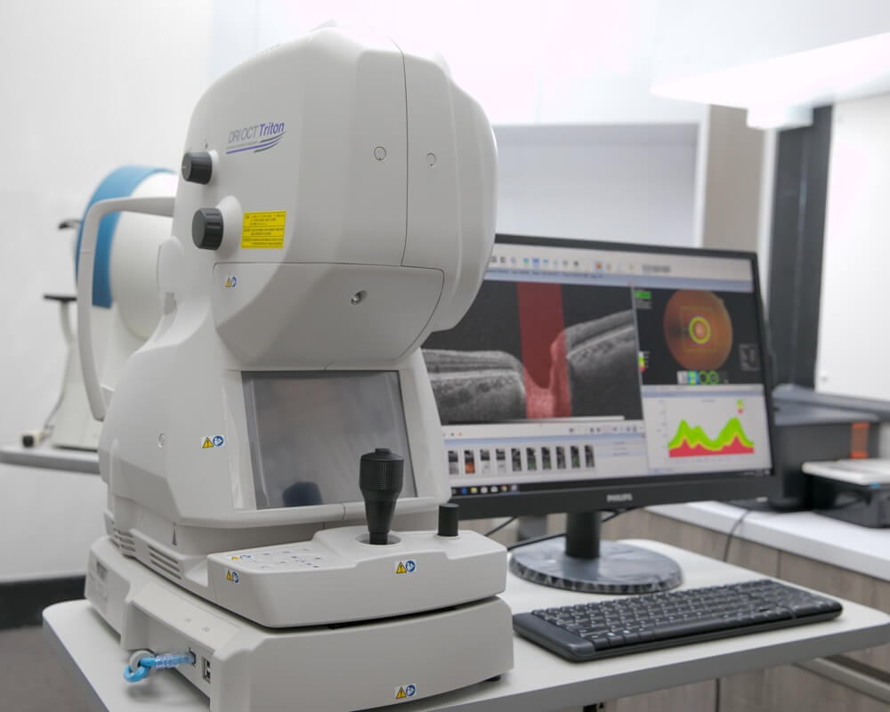
Optical Coherence Tomography or OCT is a non-invasive imaging technology that captures high resolution cross-sectional images of the eye’s posterior segment structures including the vitreous, retina, macula, optic nerves and the choroid.
OCT technology has developed rapidly and has evolved from Time-domain OCT to Spectral domain OCT and the newest Swept-source OCT.
Clinical application of OCT enables a more comprehensive observation and analysis of the physiological and pathological changes in the posterior segment of the eye such as abnormal changes caused by diabetes, glaucoma and macular degeneration.

(16) Fundus Photography
Fundus Photography
Fundus photography is a non-invasive imaging method that provides an objective view of the retina and a record for analysis, diagnosis and comparison of retinal conditions. It is a great diagnostic tool, particularly when used adjunctively with other equipment.
The fundus photo serves very well as a visual for explaining a patient’s normal or abnormal conditions.
Retinal blood vessels, optic nerves and the macula can be observed clearly in the picture taken centrally or in the mid-periphery and are extremely useful for the diagnosis and follow up of ocular diseases such as glaucoma, macular degeneration and diabetic retinopathy.

(17) Diabetic Retinopathy Screening
Diabetic Retinopathy Screening
There are over 700,000 people in Hong Kong diagnosed with diabetes, almost 10% of the total population.
High blood sugar levels cause changes in the tiny blood vessels in our organs and may eventually damage the heart, nerves, kidneys or the eyes.
Diabetes can affect the entire eye and most of them are sight threatening. They include diabetic retinopathy, maculopathy, cataract, iris neovascularization and glaucoma. As vision impairment can often be prevented with early detection of diabetic retinopathy, routine eye exams are essential for patients diagnosed with diabetes.
Diabetic retinopathy screening includes the following procedures:
- Medical history taking and complaints
- Visual function tests
- Intraocular pressure measurement
- Anterior segment examination: eyelid and annex, tears, conjunctiva, cornea, iris, crystalline lens
- Posterior segment examination: vitreous, retina and macula
- Fundus photo taking
- Optical coherence tomography
- Analysis and explanation of clinical test findings
- Suggestion of appropriate referrals or management plans according to the severity of diabetic retinopathy, macular edema, retinal hemorrhage, and abnormal blood vessels.
Primary eye care examination not only ensures that you have clear and comfortable vision but also maintains that your visual system stays healthy and functional: a key to preventive eye care.

(18) Colour Vision Test
Colour Vision Test
Colour vision is our ability to detect colours.
When we are unable to differentiate between colours, we have colour deficiency or colour blindness. If we cannot tell red from green, for example, we have red green colour defects. It is extremely rare to have people who is completely colour blind
Colour deficiency is usually inherited and affects males more but it can also be caused by eye diseases such as glaucoma, diabetic retinopathy, macular degeneration and stroke or long-term use of some medications.
Two common colour vision tests:
- Pseudoisochromatic plates for colour vision screening (e.g. Ishihara Colour Test).
- Arrangement test for types of colour defect in which a certain number of colour buttons have to be arranged in the correct order from a pilot button (e.g. Farnsworth D-15 Test).

(19) Eyewear Prescribing
Eyewear Prescribing
A good pair of spectacles must meet the following criteria:
- Appealing
- Accurate prescriptions
- Accurate optical centres
- Good optical quality
- Appropriate lens thickness
- Accurate alignment of pupil centre heights (multifocal or aspheric lenses)
- 100% UV protection, appropriate blue light filter, anti-reflection, anti-smear, scratch resistance, impact resistance, etc.
- Light, comfortable and adjustable
- Safe
Our optical lenses are supplied by major local and international lens suppliers:
- Essilor (依視路)
- HOYA (豪雅)
- Zeiss (蔡司)
- Rodenstock (萊敦司德)
- Swisscoat (瑞士寶)
- Hong Kong Optical Lens (明達)
- Polylite (寶利徠)
Refractive indices available:1.5, 1.53, 1.56, 1.59, 1.6, 1.61, 1.67 and 1.74
Lens types: Single vision for short-sightedness, long-sightedness, astigmatism and reading, bifocal, multifocal, transitions, mirror coated, tinted, polarized and sunglasses
A comprehensive eye exam is recommended for everyone before recommendations can be made based on your refractive powers, visual needs, professional or work requirements, life style and ocular health.

(20) Contact Lens Fitting
Any contact lens must fulfill all the following criteria:
- Clear vision
- Comfortable
- Safe
In our centre, we prescribe and fit the following types of contact lenses:
- Hydrogel and silicone hydrogel soft contact lenses
- Rigid Gas Permeable Lenses
- Daily wear contact lenses
- Continuous wear lenses that are worn continuously for more than one day
- Spherical lenses that correct for short or long-sightedness
- Toric lenses that correct for astigmatism
- Multifocal lenses for people who need corrections for both distance and near
- Monovision: one eye corrected for distance and the other for near
- Disposable lenses: daily, weekly, biweekly or monthly
- Conventional soft lenses that are worn daily and replaced in 6 months
- Colour contact lenses that change the appearance of the colour of your eyes
We also cater for people with special needs or conditions:
- Eyes that are sensitive, dry or easily fatigued
- Mainly for outdoor activities
- Work indoors under air-conditioning
- Needs longer wearing hours
- Occasional wearers
- Highly astigmatic
- Presbyopic (people over age 40 and require reading corrections)
- Cosmesis: different iris colours or bigger iris appearance)
As everyone is different in terms of their ocular conditions, corneal curvatures, iris diameters, tears quality, refractive errors, visual needs, life styles and working environment, anyone who wants to wear contact lenses should have their eyes properly examined before professional recommendations can be made.
To ensure long term safety, regular aftercare is essential in maintaining ocular health, clear vision and comfort.


(21) Multifocal Contact Lens Fitting
Multifocal Contact Lens Fitting
If you are over the age of 40 and beginning to find near prints a bit blurry and difficult to focus with your current contact lenses, then you may start to have a condition medically known as ‘presbyopia’ which literally means ‘old eye’. Or you might have already given up contact lenses for years for the same reason, then here is good news!
The new generation multifocal contact lenses will help solve your vision problems for both distance and near, whether you are shortsighted, longsighted or you never require distance corrections before.
Different from single vision contact lenses, these multifocal contact lenses have 3 zones at the centre of the lens corresponding to distance, intermediate and near vision. Depending on the design, the lens can be centre distance or centre near. The eye will receive all three different images before it transmits them to the brain which through integration will in turn provide the vision for a particular distance required. For example, when we read, the brain will process the near zone image for us so that we can see at near.
(Source: Cooper Vision 酷柏光學)
Current multifocal contact lens designs are quite well developed with satisfactory results. They are easy to use and most people will adapt within 2 weeks.
There are numerous designs, materials, wearing modalities available on the market. We are anticipating a major breakthrough in this category in the not so distant future. Based on individual ocular conditions and visual requirements, we will strive to fit the most appropriate multifocal contact lenses for you.

(22) Health Care Voucher
Health Care Voucher
The Elderly Health Care Voucher Scheme was initially launched on 1 January 2009 to try out a new concept of enhancing the provision of primary care service for the elderly aged 70 or above. The Scheme aims to supplement existing public healthcare services (e.g. General Out-patient and Specialist Out-patient Clinics) by providing financial incentive for elders to choose private healthcare services that best suit their health needs, including preventive care
The Government has lowered the eligibility age for the Scheme from 70 to 65 with effect from 1 July 2017.
In 2018 and 2019, apart from the annual voucher amount of $2,000, each eligible elder was also provided with an additional voucher amount of $1,000 on a one-off basis on 8 June 2018 and 26 June 2019 respectively while the accumulation limit of the vouchers was increased to $8,000 with effect from 26 June 2019 as a regular measure.
Besides, in view of the outcome of a review on the Scheme in 2019, a cap of $2,000 every two years on the voucher amount that can be spent by each elder on optometry services has been introduced with effect from 26 June 2019 to encourage the use of vouchers on different primary healthcare services.
Primary eye care examination provides a comprehensive eye examination for the elderly and delivers preventive and rehabilitative eye care services with respect to their refractive needs and ocular health management.

(23) Eyesight Test Certificate – Pleasure Vessel Operator and Local Vessel Coxswain
Eyesight Test Certificate – Pleasure Vessel Operator and Local Vessel Coxswain
Our centre provides eyesight tests for Pleasure Vessel Operators and Local Vessel Coxswains according to the standards required by the Marine Department and issues the Eyesight Test Certificate if the applicant meets the standards.

Excellent Facility
State of the Art Equipment:

Axial Length Scan

Optical Coherence Tomography

Corneal Topography

Digital Bio-microscope

Visual Field Tester

Fundus Camera
BOOK AN APPOINTMENT
Our comprehensive Eye Examination is Suitable for All Family Members.